Tibiofibular Joint Sprain
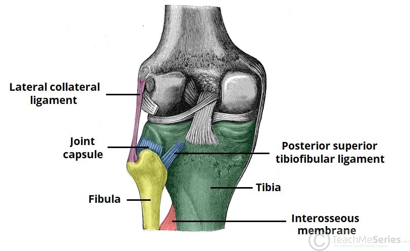
Severity: Mild to Severe, depending on location and degree of injury
Typical recovery time: Most proximal injuries resolve within a few weeks with conservative treatment. Distal tibiofibular syndesmosis sprains typically take longer than lateral ankle sprains, often 6–12 weeks, and significant instability may require surgical fixation
Can I keep running? No in the acute phase. Running places rotational and impact forces directly risks progressing the injury
What's Actually Happening: The tibia and fibula are connected at two joints: the proximal tibiofibular joint (PTFJ) near the outer knee and the distal tibiofibular syndesmosis just above the ankle. Both serve to allow small controlled movements that coordinate loading between the knee and ankle during running. The PTFJ is a small plane synovial joint designed to rotate slightly to accommodate rotational stress at the ankle during dorsiflexion. It is heavily supported by surrounding ligaments and rarely injured, accounting for only around 1% of all knee injuries. When the PTFJ is sprained or dislocated it causes outer knee pain that can be confused with IT band syndrome, lateral meniscus injury, LCL sprain, or posterolateral corner injury. The distal tibiofibular syndesmosis is more commonly injured during sports and accounts for 11–17% of all ankle sprains in athletic populations, typically from a forced external rotation or dorsiflexion of the foot. These are sometimes called high ankle sprains since the pain sits above the lateral ankle rather than over it. Both joints can also develop ganglion cysts that compress the nearby common peroneal nerve, causing tingling, numbness, or foot drop, which is worth knowing as an uncommon complication of PTFJ injury.
How You Know It's This (Symptoms):
Proximal tibiofibular joint:
Pain around the fibular head on the outer knee, often with clicking, shifting, or feelings of instability
Tenderness directly over the fibular head when pressed
Pain worsening with squatting, pivoting, hill running, and kneeling
Occasional tingling or numbness down the outer lower leg if the nearby common peroneal nerve is irritated
Symptoms that overlap with IT band syndrome and lateral knee problems, making accurate diagnosis important
Distal tibiofibular syndesmosis (high ankle sprain):
Pain localized above the lateral ankle rather than over it, distinguishing it from a standard lateral ankle sprain
Pain that is sharp and localized at the front or back of the distal fibula just above the ankle joint
Tenderness over the anterior tibiofibular ligament just above the ankle
A squeeze test, squeezing the tibia and fibula together at mid-calf, reproduces the pain at the syndesmosis and is one of the most reliable clinical signs
Significant difficulty bearing weight in more severe cases
What To Do Right Now:
Stop running and all activities involving rotational loading of the ankle or knee
Ice the affected area for 15–20 minutes several times daily in the first 48 hours
For distal syndesmosis injuries, avoid bearing weight if the squeeze test is positive or if walking causes significant pain
Seek medical evaluation for any suspected tibiofibular injury since the degree of instability determines the treatment approach and conservative versus surgical management decisions are based on clinical and imaging findings
When to See a Professional:
Any outer knee pain with clicking, fibular head tenderness, or instability that doesn't resolve within a week or two warrants assessment to rule out PTFJ injury alongside more common lateral knee problems
MRI is the most appropriate imaging tool for PTFJ soft tissue injuries. X-rays are needed to rule out fibular fracture and assess for syndesmotic widening in distal injuries
How to Treat It:
Conservative management for most cases: Most tibiofibular joint sprains without significant instability resolve with conservative treatment. The standard approach includes activity modification, physiotherapy, manual therapy, and a staged return to running.
Physiotherapy: Treatment focuses on load modification, taping, bracing, manual therapy, strength work, balance training, and a staged return to running. For the PTFJ specifically, proprioception and lower limb control exercises are important components since joint instability is a significant contributor to ongoing symptoms. For syndesmosis injuries, rehabilitation follows a similar progression to lateral ankle sprain rehabilitation but with a longer timeline.
Taping and bracing: Taping the tibiofibular joint provides external support and reduces abnormal movement during rehabilitation and return to running. For syndesmosis injuries, a documented case report showed that a player taped before every practice with a lateral ankle restraint and circumferential strips proximal to the distal tibiofibular joint for the remainder of the season experienced no re-injury, persistent pain, or loss of function.
Manual therapy: Joint mobilization by a physiotherapist can restore normal joint mechanics and reduce pain, particularly for PTFJ stiffness or mild instability.
Surgery for instability: Significant proximal tibiofibular joint instability or dislocation that doesn't respond to conservative treatment may require surgical reconstruction, with a modified ACL reconstruction protocol used as a guideline given the lack of specific evidence-based protocols for this rare condition. For distal syndesmosis injuries with frank instability, surgical fixation with screws or a tightrope device restores normal joint width.
The Recovery Plan:
Week 1–2: Stop running, ice the affected joint, and begin physiotherapy assessment to determine degree of instability. Taping or bracing during daily activity. Most patients with a tibiofibular joint injury begin to recover within a few weeks with appropriate rehabilitation.
Week 2–8: Progress strength, balance, and proprioception work. For PTFJ injuries, most cases resolve completely with conservative treatment within this window. For syndesmosis injuries, timeline is longer than a standard ankle sprain — rushing return to running before adequate healing leads to chronic instability.
Beyond 8 weeks: Gradual return to running on flat surfaces once the joint is pain-free with normal function. Continue taping during running for the remainder of the season if returning mid-season. Persistent instability or failure to improve with conservative management warrants surgical consultation.
