Spondylolysis
Severity: Moderate
Typical recovery time: A minimum of 3 months of activity restriction is the standard recommendation; with immediate functional rehabilitation, some athletes return to sport in as little as 2.5 months.
Can I keep running? No. Running and sports involving back extension must stop until the injury is confirmed healing on imaging
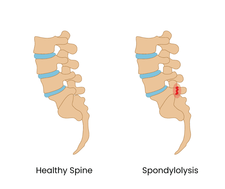
What's Actually Happening: Spondylolysis is a bone stress injury of the pars interarticularis, a small bony bridge connecting the upper and lower facet joints of a vertebra. It most commonly affects the fifth lumbar vertebra (L5) and is more prevalent in male athletes. The pars interarticularis is uniquely vulnerable to the shear forces created during repeated lumbar hyperextension movements, making sports involving repetitive back extension — including gymnastics, wrestling, and distance running with an anterior pelvic tilt — particularly high-risk. In adolescent athletes, low back pain is quite common and spondylolysis is the most common identifiable cause, meaning this is a genuinely important injury to know about for teen cross country and track athletes. Left untreated, a spondylolysis can progress to spondylolisthesis, where the fractured vertebra slips forward on the one below it, which is a more serious condition. The good news is that the prognosis with appropriate conservative management is excellent: a review of over 200 adolescent athletes treated with a standardized conservative protocol found that 98% returned to competition, with only one requiring surgical management.
How You Know It's This (Symptoms):
Focal lower back pain, typically on one side, that worsens with running and especially with back extension movements like arching the back or doing a back bend
The one-legged hyperextension test is a commonly used clinical screen: standing on the affected side leg and extending the back reproduces the pain in many cases
Pain that improves significantly with rest and returns predictably with return to running or sport
Absence of radiating leg pain or neurological symptoms in most cases, which helps distinguish it from disc-related problems
Gradual onset over weeks of increased training rather than a single traumatic event
What To Do Right Now:
Stop all sports activity and running immediately
Avoid any back extension movements including arching the back, gymnastics-type movements, and certain core exercises like supermans
See a doctor as soon as possible, since diagnosis requires imaging. Plain X-rays may appear normal in early or stress reaction stages and MRI is the more sensitive early tool, with CT providing the most detailed view of bony healing
Do not attempt to manage this one conservatively at home without proper imaging and diagnosis — knowing the specific stage and severity of the fracture guides the treatment plan
When to See a Professional:
This injury requires professional evaluation from the moment it's suspected
An MRI is the recommended first imaging tool for adolescents since it avoids radiation and is sensitive for early stress reactions before a visible fracture line appears
Spondylolysis that progresses to spondylolisthesis, where the fractured vertebra slips forward, requires specialist orthopedic management and changes the treatment approach significantly
Surgery is indicated only when patients remain symptomatic despite more than one year of nonoperative management
How to Treat It:
Activity restriction and bracing: The traditional standard treatment involves complete sports cessation and a thoracolumbosacral orthosis (TLSO) brace for a minimum of 3 months. In a study of over 200 adolescent athletes treated with this protocol alongside an external bone stimulator, 98% returned to competition. However, it remains unclear whether the brace itself or the activity restriction contributes more to outcomes, and there is emerging interest in more immediate rehabilitation-based approaches.
Immediate functional progression (emerging approach): A study proposing an immediate functional progression program found that young athletes were able to make a full return to sport approximately 2.5 months from diagnosis, challenging the traditional model of extended rest. This approach begins rehabilitation immediately rather than waiting for symptom resolution, aiming to treat the movement impairments that led to the injury, avoid muscle atrophy from rest, and improve quality of life during recovery. While promising, this approach is still being investigated and should only be pursued under close clinical supervision.
External bone stimulator: Used alongside bracing in many treatment protocols to promote bony healing of the pars defect. While evidence for its specific contribution is limited, it was included in the protocol that achieved 98% return to competition and is commonly prescribed in clinical practice.
Physical therapy and core stabilization: Although there are no randomized controlled trials specifically investigating rehabilitation for spondylolysis in adolescent athletes, physical therapy is consistently recommended and clinical outcomes are excellent. Core stabilization targeting the deep stabilizers of the lumbar spine reduces the shear forces on the pars interarticularis during movement, addressing the mechanical root cause of the injury.
Corticosteroid injection: Occasionally used for pain management in athletes with significant ongoing pain that is preventing engagement with rehabilitation, guided by imaging to target the pars defect specifically.
Surgery: Direct pars repair is reserved for cases with documented non-union after more than one year of conservative management and a clear desire to return to sport. It involves debridement of the fibrous tissue at the fracture site, bone grafting, and fixation. Surgical outcomes are generally good for fractures above L5, though the non-union rate is higher at the L5 level due to shear forces at that segment.
The Recovery Plan:
Month 1–3: Complete cessation of sports and running. Bracing with a TLSO as directed by your physician. External bone stimulator if prescribed. Imaging at 3 months to assess healing on CT scan.
Month 3–6: Return to sport is guided by both symptom resolution and imaging findings. If CT confirms adequate healing and symptoms have resolved, a graduated return to physical therapy and then sport-specific training begins. An immediate functional progression program may begin earlier under close clinical supervision in appropriate cases.
Return to running: Gradual return to running begins only once cleared by a physician based on clinical and imaging findings. Begin with easy flat running, avoid back extension loading, and progress mileage slowly. The full return to competitive running typically occurs between 3 and 6 months from diagnosis with appropriate management.
